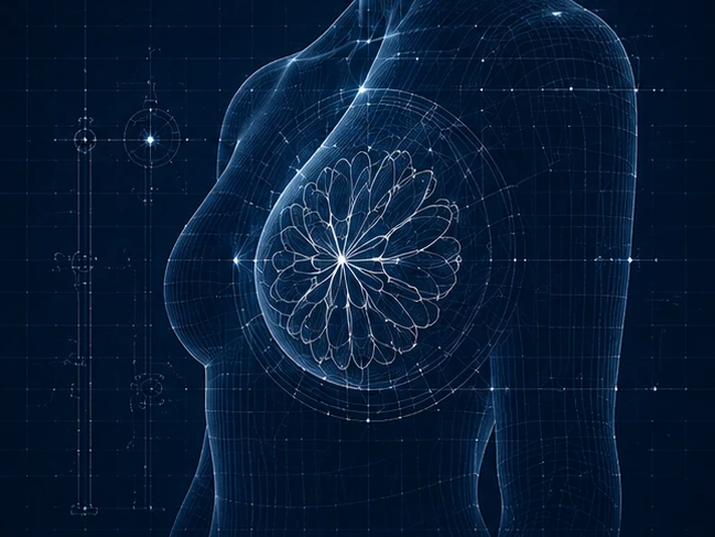
Thermography

Seeing What Other Tools Can't

Traditional breast screening answers one question: What is present?

Thermography answers a different question: What is happening?

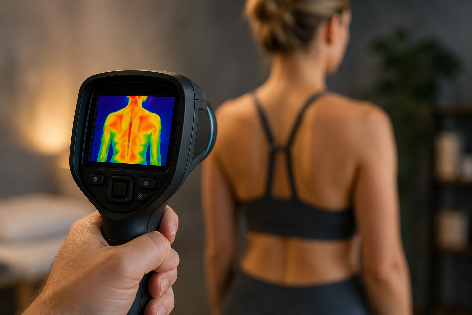
Infrared Thermography uses an FDA-registered medical-grade infrared camera to detect subtle variations in heat radiating from the body’s surface. It identifies early metabolic and circulatory changes in tissue — changes that occur as breast tissue becomes more active, inflamed, or disrupted, often well before a structural change becomes visible on imaging.

The principle is straightforward: as metabolic activity increases in an area of concern, so does local circulation and heat. The infrared camera detects these heat patterns with a sensitivity that no other screening tool can match at the functional level.

No radiation. No compression. No contact of any kind.

How It Compares to Mammography

Mammography uses X-rays to capture structural information — it answers the question of what is present by imaging dense tissue. It has approximately 80% sensitivity in women over 50, which means it misses roughly 20% of cancers in this group. In women under 50, particularly those with dense breast tissue, sensitivity is lower still. Dense tissue, implants, fibrocystic breasts, and hormone replacement can all limit mammography’s effectiveness.

Thermography detects physiological changes in tissue rather than structural changes. It has approximately 90% sensitivity across all age groups. It is particularly effective for dense breast tissue (where mammography sensitivity is reduced by up to 40%), younger women, fibrocystic breasts, women with implants, and those who have had breast reduction or lumpectomy.

It is important to understand that neither mammography nor thermography diagnoses cancer. Only a biopsy can determine whether a questionable area is malignant. Thermography is designed to complement traditional screening — not replace it. Used alongside mammography and a thorough clinical evaluation, it creates a more complete picture than either tool provides alone.

Research has shown that thermography may detect pre-cancerous metabolic changes 5 to 8 years earlier than traditional structural screening methods.

What to Expect

The thermography procedure consists of three steps and takes approximately 30 to 45 minutes.

Step 1 — Preparation: You complete your Breast History Form and arrive 15 minutes before your appointment.

Step 2 — Equilibration: You disrobe from the waist up and rest comfortably in a temperature-controlled room (approximately 70°F) for 15 to 30 minutes, allowing your body’s temperature to equilibrate with the room. This step is essential for accurate imaging.

Step 3 — Imaging: Your provider takes 3 to 8 infrared images from approximately 2 to 4 feet away. After the initial images, you will be invited to place your hands in cold mittens for 60 to 90 seconds. This cold challenge alters blood flow — healthy tissue responds by redirecting blood to the core, while inflamed, infected, or abnormal tissue does not. The contrast between responding and non-responding tissue provides additional diagnostic information. Images are then repeated and sent for professional interpretation.

Results are provided via report, email, phone call, or at a follow-up visit depending on your situation.

Preparing for Your Appointment

Accurate thermography results depend on careful preparation. Please follow these guidelines:

24 hours before: Avoid all physical stimulation or treatment of the area to be imaged — chiropractic care, massage, physical therapy, acupuncture, saunas, heating pads, ice, analgesic creams, laser or ozone therapy, sun exposure, or dry needling. Do not shave the armpits or chest area.

12 hours before: Avoid external breast prosthesis.

4 hours before: Avoid caffeine, alcohol, hot baths or showers, rigorous exercise, nicotine, and vasoactive medications if possible.

1 hour before: Do not bathe or shower. Nursing mothers should not nurse within one hour of the appointment.

Examination is optimal when not performed between the 6th and 13th day of the menstrual cycle. If you have had a sunburn, fever, or skin burn, please reschedule. Inform us if you have had chemotherapy or radiation within the last two months.

Stop Guessing. Start Understanding.

What It Helps

Thermography is particularly valuable for women with dense breast tissue, those seeking a complementary view alongside mammography, women with implants or previous breast surgery, younger women for whom mammography is less effective, pregnant or lactating women who need a radiation-free option, and women who want earlier visibility into metabolic and circulatory patterns in breast tissue.

10 Tips for Overcoming Joint Pain

Enter your information below to learn how we've helped thousands treat the root source of their pain and helped them truly heal by triggering the body's innate healing system.

You have Successfully Subscribed!